


2026 Annual Meeting of the
International Anaplastology Association

Dear Colleagues,
It is with great pleasure that I invite you to Baltimore, Maryland for the 39th Annual Meeting of the International Anaplastology Association! This year’s conference will be held from July 8–11, 2026, at the Armstrong Medical Education Building on the East Baltimore campus of Johns Hopkins School of Medicine and Hospital. Accommodations are available at the Hyatt Harbor Place Hotel, located in the vibrant Inner Harbor neighborhood—just a short, chartered bus ride from the conference venue.
Our meeting offers a unique opportunity to bring together clinicians, rehabilitative specialists, physicians, and a wide range of medical professionals to share insights and advance the care we provide to our patients.

IAA2026 CONFERENCE THEME:
The theme of this year’s gathering Outcomes, Transformation, & Resilience centers on the profound impact of clinical anaplastology through the lens of our patients. While the pursuit of a “perfect” prosthesis is often constrained by the realities of replicating absent or malformed anatomy, our impact as clinicians lies in deepening our understanding of anatomy, material science, retention methods, and the artistic and technical skills that elevate our practice. A well-crafted prosthesis can transform lives—enhancing daily function, social engagement, protection, restoration, and self-confidence, while helping patients embrace a “new normal.”
We invite presentations that explore the transformative power of facial, ocular, and body prosthetics—not only as medical devices but as instruments of resilience and hope. Hands-on workshops will offer immersive learning in digital workflows, cutting-edge implant retention systems, and anatomical lectures to strengthen clinical knowledge and skills.
Baltimore, affectionately known as Charm City, is a historic and culturally rich destination. The conference site is just steps from the iconic dome of Johns Hopkins Hospital and near the renowned Department of Art as Applied to Medicine, founded in 1911 and celebrated for its graduate programs in Medical and Biological Illustration and Clinical Anaplastology.
Beyond the conference, Baltimore offers a dynamic mix of experiences—from world-class institutions like the Walters Art Museum and Baltimore Museum of Art to cozy, stylish bars and restaurants. The Hyatt Harbor Place Hotel, nestled in the Harbor East neighborhood, is within walking distance of Little Italy, where you can enjoy a delicious pasta dinner followed by a cannoli. The hotel also provides easy access to charming districts like Fells Point, and its location by the Inner Harbor makes it perfect for a morning jog or a scenic Water Taxi ride to Fort McHenry, birthplace of our National Anthem. Baltimore promises a memorable backdrop for our gathering.
Warm regards,
Juan Garcia, MA, CCA
Vice President, International Anaplastology Association
We hope that you'll plan to join us in Baltimore in JULY 2026

Anaplastology is a specialized healthcare field that provides patient-specific, restorative prostheses for patients whose anatomy is absent, altered, or lost due to injury, disease, congenital origin, or other wholeness-limiting conditions. Customized prosthetic rehabilitation may be an alternative treatment option to surgical reconstruction. Anaplastology services are intended to restore affected anatomical features of the body including but not limited to the face, hands, feet, or breast. The scope of the anaplastologist may also include custom medical device design applied to restorative interventions in collaboration with other medical specialties.
The IAA promotes quality patient care by supporting the development of best practices in anaplastology through educational conferences, networking, publication, and advocacy opportunities.


IAA2026 SCHEDULE OVERVIEW
July 8-11, 2026
JULY 8th DAY ~ Pre-conference Paid Workshops
Pre-conference Courses will be held on JULY 8th. These events have limited capacity. To register, select the pre-conference event(s) you wish to attend on your registration form to secure your place.
JULY 8th NIGHT ~ Welcome Reception
Hosted reception at Kneads Bakery from 6:30 to 8:00 pm for conference attendees and vendors.
JULY 9th &10th DAY ~ Scientific Sessions
Scientific Sessions will take place at the West Lecture Hall at the Armstrong Medical Education Building
JULY 9th DAY ~ Mid-conference Paid Workshop
A Mid-Conference Course will be held on JULY 9th. This event has limited capacity. To register, select the mid-conference event on your registration form to secure your place.
JULY 10th NIGHT ~ IAA Banquet, Awards, & more . . .
Join us at 6:30 pm at Chiapparelli's restaurant in Little Italy! Buy your tickets in advance to save your spot!
And then meet back at the Hyatt Place Baltimore/Inner Harbor hotel at 8:30 pm for Post-Banquet Awards / Oto Art Event.
JULY 11th DAY ~ Post-conference Paid Workshops
Post-conference Courses will be held on JULY 11th. These events have limited capacity. To register, select the post-conference event(s) you wish to attend on your registration form to secure your place.
IAA2026 CONFERENCE
IMPORTANT INFO
-
This year's conference will feature engaging lectures, hands-on demonstrations, interactive group activities, free vendor presentations, planned social events, and optional fee-based workshops designed to enhance learning and professional connection.
-
All conference events will be held in-person in Baltimore, Maryland, USA on July 8-11, 2026
-
Conference Venue - Armstrong Medical Education Building on the East Baltimore campus of Johns Hopkins Medical School and Hospital.
-
Host Hotel – Hyatt Place Baltimore/Inner Harbor. Make your hotel reservations now for our 2026 IAA Conference! Click this link to make your reservation today.... Book IAA Hotel Rate
-
Transportation - Due to the distance between the hotel and conference venue, walking is not recommended. Convenient private Shuttle Busses will be provided at scheduled times. Guests who miss the shuttle are encouraged to use taxis or ride-share services (Uber/Lyft) to the Armstrong Medical Education Building (1600 McElderry St., Baltimore), located directly across from the Johns Hopkins Hospital Outpatient Center (601 N. Caroline St., Baltimore) and the hotel.
-
Social Events - include the IAA Banquet in Little Italy (additional fee) and after-dinner event at the Hyatt Place Baltimore/Inner Harbor hotel, tours of Johns Hopkins Facial, Eye & Body Prosthetics Clinic and the Clinical Anaplastology Graduate Program at the JHU Department of Art as Applied to Medicine.

REGISTRATION NOW OPEN!
Interested in becoming a member?
Click here to be redirected to the IAA Membership Application Form.
EARLY BIRD DEADLINE EXTENDED THROUGH 5PM EST MAY 25th!


IAA2026 WORKSHOPS
Check out all the educational options!
Pre, Mid, and Post Conference Workshops available for purchase... sign up early as space is limited!
Workshop 1 1:30 - 4:30pm
Surgical Anatomy Review
Mark Fisher, MD
& Johns Hopkins surgical residents
Course offers a unique opportunity to review relevant surgical anatomy of midfacial, temporal bone and hand regions with practicing surgeons. Teaching resources include prosected cadaver specimens, plastinated specimens and lectures featuring medical illustrations.
Cost - $300.00 USD Capacity - 24 attendees
Workshop 2 1:00 - 5:00pm
ZBrush Workflows
for Anaplastologists
Juan Garcia, MA, CCA, Andrew Etheridge, MFA, CCA, CFo, CFm, C.Ped, BCO, BADO &
Paul Gaboury, Senior ZBrush Product Development Manager
Course begins with a one-hour live Zoom demonstration led by Paul Gaboury, technical expert from Maxon ZBrush, includes a showcase of the iPad version of ZBrush. Followed by a series of hands-on exercises with Juan Garcia, CCA, where participants can follow along.
Course held in Mac Computer Lab equipped with Maxon ZBrush and Wacom pressure sensitive pens. Teaching covers common workflows such as importing/aligning 3D scan or CT data, methods for cropping/mirroring/Boolean operations/achieving thin margins/shell creation, and preparing models via Scale Master, Decimation Master and 3D Print Hub for final 3D printing.
Cost - $300.00 USD Capacity - 18 attendees
Mid-Conference Workshop
Thursday, July 9, 2026
Workshop 3 3:30 - 6:00pm
BioComp AHEAD Osseointegrated Retention Hands-On Session
Maarten de Jong, MA, Anaplastologist & BioComp Implant company representatives
Course content includes lectures on system components, pre-surgical planning and post-surgical considerations with an opportunity for hands-on surgical simulation. Course taught using temporal (auricular region) and midface (eye and nasal region) bone-like drillable models. In addition, prosthesis design considerations will be discussed.
Cost - $250.00 USD Capacity - 18 attendees
Post -Conference Workshops
Saturday, July 11, 2026
Workshop 4 8:00 - 10:30am
Southern Implants Osseointegrated Retention Hands-On Session
Southern Implant company representatives,
University of Illinois (UIC) & Johns Hopkins
faculty physicians
Course content includes lectures on system components, pre-surgical planning and post-surgical considerations with an opportunity for hands-on surgical simulation. Course taught using temporal (auricular region) and midface (eye and nasal region) bone-like drillable models. In addition, prosthesis design considerations will be discussed.
Cost - $250.00 USD Capacity - 18 attendees
Workshop 5 8:00 - 10:30am
Ocular Painting
using Light-Cured Acrylic
Marie Drennan, BCO, BADO
& Heather Meszaros, BCO
Hands-on opportunity to use of Palaseal light cured acrylic dental sealant as a painting medium. Relevant eye anatomy will be covered and an ocular prosthesis with a blue iris will be painted during the workshop. Each participant provided with supplies and tools needed for the workshop, including a previously prepared ocular shape with an embedded black iris button.
Cost - $200.00 USD Capacity - 24 attendees
Workshop 6 11:00 am – 1:00 pm
Prosthetic-Driven Implant Planning using BlueSkyPlan & Digital Workflows for Anaplastology Cases
Ghassan Sinada, DDS, MBA
Paula Sauerborn, MA, CCA
Juan Garcia, MA, CCA
& Maarten de Jong, MA, Anaplastologist
This demonstration workshop introduces a team approach to hybrid and digital prosthesis workflows using BlueSkyPlanV5 AI-driven modules for automated bone segmentation and alignment of an imported proposed prosthesis envelope (STL) based on the patient’s unique anatomy. This comprehensive workshop covers the digital workflow click-path from data import and precise implant positioning with clinical verifications, to design of a patient-specific implant surgical guide ready for export and subsequent 3D printing.
Following the workshop, registrants will be provided with access to the DICOM and STL training data sets, as well as BlueSky Bio Export Coupons ($250 value), allowing for independent practice and implementation of presented workflows.
Cost - $250.00 USD Capacity - 170 attendees
Pre -Conference Workshops
Wednesday, July 8, 2026

IAA2026 PRESENTATIONS
Featured & Invited Speakers
(click on speaker's headshot to read their BIO)
WST Featured Speaker
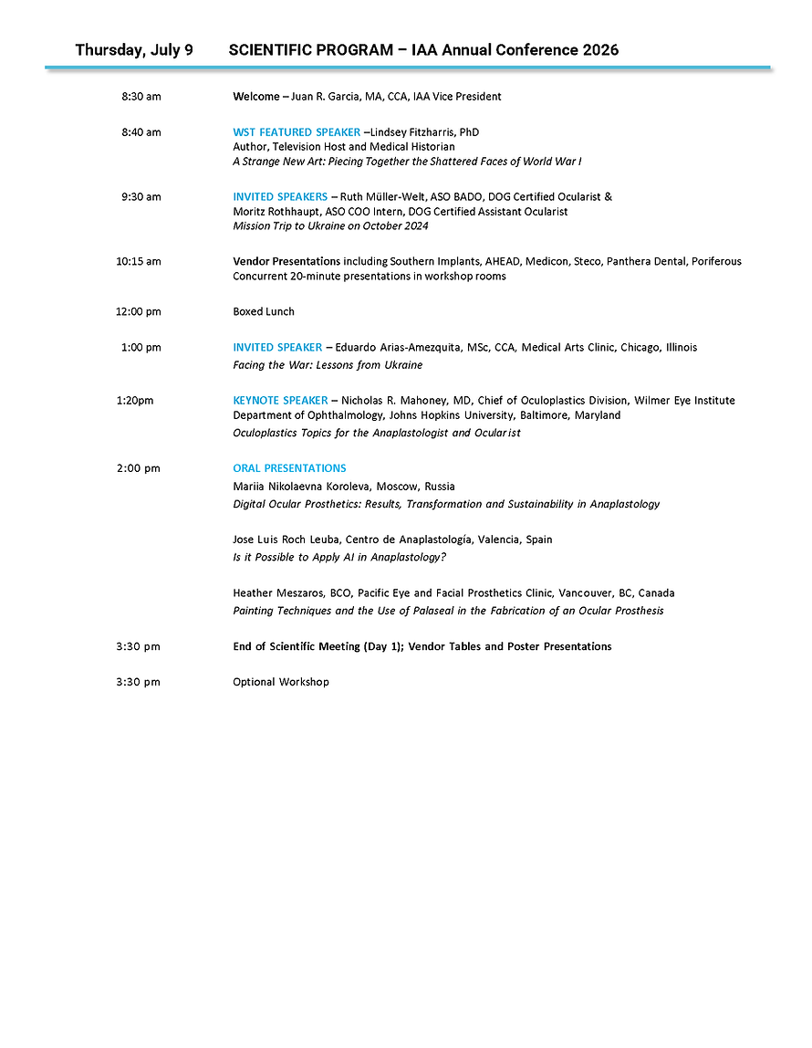
A Strange New Art: Piecing Together the Shattered Faces of World War I

From the moment the first machine gun rang out over the Western Front, one thing was clear: mankind’s military technology had wildly surpassed its medical capabilities. Bodies were battered, gouged, hacked, and gassed. The First World War claimed millions of lives and left millions more wounded and disfigured. In the midst of this brutality, however, there were also those who strove to alleviate suffering. One such individual was the pioneering plastic surgeon Sir Harold Gillies, whom together with an extraordinary interdisciplinary team that included dentists, artists, and mask-makers, taught himself and others how to repair the burned and broken faces of the injured soldiers under his care. At its core, this is a story about how medicine and art can merge, and of what courage and imagination can accomplish in the presence of relentless horror.
Featured Speaker
Oculoplastics Topics for the Anaplastologist and Ocularist

Nicholas Mahoney, MD
Professor of Opthamology,
Johns Hopkins University School of Medicine
Baltimore, Maryland, USA
This presentation will explore key topics in oculoplastics relevant to the anaplastologist and ocularist. It will include an overview of eye removal procedures and current trends, as well as common eyelid malpositions, their underlying causes, and management strategies.
The session will also review modern techniques used to restore orbital volume in the anophthalmic socket, with a focus on approaches that support both functional and aesthetic outcomes.
Featured Speaker
ENT Constructs: Sculpting Noses and Ears from Living Tissue or Silicone

Kofi Boahene, MD
Professor of Otolaryngology,
Johns Hopkins University School of Medicine
Baltimore, Maryland, USA
Reconstruction of the nose and ear demands a precise balance of structure, aesthetics, and durability. This presentation explores “ENT constructs” as a design-driven approach to shaping nasal and auricular frameworks using either living tissue or silicone.
A key focus is the collaboration with anaplastologists, whose expertise in creating surgical guides enhances precision in reconstructing form using autologous tissue. This interplay between surgical technique and prosthetic design allows for more predictable, patient-specific outcomes.
We will highlight principles of material selection, structural design, and soft tissue integration, along with strategies to optimize contour and longevity. Ultimately, this approach reframes reconstruction as a collaborative process—where biology and design converge to restore both form and identity
Featured Speaker
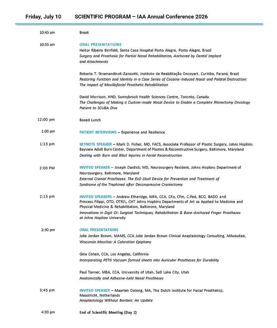
Dealing with Burn and Blast Injuries in Facial Reconstruction

Mark Fisher, MD FACS
Associate Professor of Plastic Surgery,
Johns Hopkins University School of Medicine
Baltimore, Maryland, USA
The hallmark of anaplastology is the achievement of the highest possible reconstructive outcome with the lowest morbidity. This aim however can be challenging when patient specific factors create a hostile environment for our tools. What do we do if there is abundant scarring? How do we deal with contracting scars arising from massive trauma, burns, or past infections? In this session we will explore patients who have suffered burn, blast, and infection resulting in reconstructive needs and how these mechanisms influence our recipient site preparation and ultimate reconstruction whether using autologous or anaplastic approaches.
Invited Speaker
External Cranial Prostheses:
The ExO-Skull Device for Prevention and Treatment of Syndrome of the Trephined after Decompressive Craniectomy

Joseph Dardick, MD
Neurosurgery Resident,
Johns Hopkins Department of Neurosurgery,
Baltimore, Maryland, USA
Decompressive craniectomy (DC) is a life-saving surgery for patients suffering from malignant brain swelling secondary to conditions such as stroke and traumatic brain injury. In DC, a large piece of skull is removed to relieve intracranial pressure. Cranioplasty, surgery to reconstruct the skull, is most safely performed 3–6 months after DC. During this window, patients live with an incomplete skull and as many as 65% will experience Sunken Flap Syndrome (SFS), new and worsened neurologic symptoms that improve only after cranioplasty.
Isolated reports of external cranial prostheses (ECP) show promise for treating SFS, but no interventions have undergone rigorous evaluation for the prevention of SFS or improvement of neurologic function during the craniectomy-to-cranioplasty window. We have initiated the External post-Operative Skull prosthesis (ExO-Skull) trial to evaluate the ability of ECPs to prevent SFS, improve neurologic recovery, and decrease healthcare utilization during this critical window.
In this trial, patients receive a custom-printed ECP and are followed with serial neurologic evaluations. The ExO-Skull device is 3D printed and designed based on each patient’s specific anatomy to optimize patient comfort and isolation of the brain. To date, the ExO-Skull device has demonstrated efficacy for treating SFS in a test case as well as safety, patient/family satisfaction, and efficacy when used for SFS prevention after DC. The trial is already enrolling and will be the first to rigorously investigate a treatment for SFS in this vulnerable patient population.
Invited Speakers
Mission Trip to Ukraine on October 2024

This presentation is a review of Moritz Rothhaupt and my mission trip to
Kiev, Ukraine by the end of October 2026. We will give you an overview about our journey, the people, the patients, and the professionals we met. We were not there to fit many patients - but to give our knowledge to specialists at and around the clinic - leading to total of 151 eye prostheses made in 2025 for the soldiers.
Ruth Müller-Welt, ASO BADO
DOG Certified Ocularist, ASO Diplomate
Augenprothesen, Stuttgart, Germany

Moritz Rothhaupt, ASO
COO Intern, DOG Certified Assistant Ocularist
Augenprothesen, Stuttgart, Germany
Invited Speaker
Facing the War: Lessons from Ukraine

Eduardo Arias-Amezquita,
MSc, CCA
Medical Arts Clinic
Chicago, Illinois, USA
Russia’s full-scale invasion of Ukraine in 2022 created one of the most severe injury crises in modern conflict, including tens of thousands of amputations and a rise in complex facial trauma. This talk examines our wartime experience planning and treating complex facial injuries among soldiers in Ukraine. It will demonstrate how we responded through innovative surgical facial reconstruction and anaplastology rehabilitation, in collaboration with Ukrainian and American surgeons under the guidance of the international NGO Razom for Ukraine, during the American Academy of Facial Plastic & Reconstructive Surgery (AAFPRS) Face-to-Face medical mission in March 2026.
Drawing on clinical experience, this talk covers three interconnected areas: the production of same-day ocular devices for oculoplastic surgical reconstructions; the surgical and technological response to combat-related facial injuries, including planning and placement of BioComp® AHEAD craniofacial implants; and the urgent need for facial prostheses, where demand far outstrips current capacity.
This talk also addresses the human dimension: the psychological burden of working under fire, the profile of patients—predominantly young, active men—and the challenge of building sustainable local expertise under wartime conditions. Patient stories ground the clinical and technical details in lived reality. Ukraine is not merely absorbing foreign aid—it is generating new knowledge, new techniques, and new technologies. The lessons being learned here will shape a humanitarian mindset for years to come.
Invited Speakers
ASO Program and NEBO Credentials for Ocularistry

Jean Thompson,
BCO, BADO, FASO
Thompson Ocular Prosthetics, Inc.
San Antonio, Texas, USA
For those who are interested in becoming a Board Certified Ocularist, this lecture will provide an overview of the educational requirements and pathways available. The National Examining Board of Ocularists, College of Ocularistry, American Society of Ocularists and International Academy of Ocularists, will all be discussed and how they contribute towards the education process of becoming certified. Visuals of cases typically seen in an ocularist practice will be included in this presentation.
%20Headshot.jpg)
Marie Drennan, BCO, BADO
Pacific Eye & Facial Prosthetics Clinic
Vancouver, British Columbia, Canada
Invited Speakers
Innovations in Digit OI: Surgical Techniques, Rehabilitation & Bone-Anchored Finger Prostheses at John Hopkins University

Andrew Etheridge,
MFA, CCA, CFo, CFm, C.Ped, BCO, BADO
Instructor of Art as Applied to Medicine,
Johns Hopkins University School of Medcine,
Baltimore, Maryland, USA

This lecture presents a detailed case report demonstrating a multi-disciplinary approach to bone-anchored digit rehabilitation. The presentation outlines the patient’s rehabilitation journey, including the development and implementation of an axial loading and traction protocol designed to promote successful osseointegration. In addition, the case highlights patient-specific orthotic and prosthetic design strategies developed to optimize functional outcomes of the hand.
The results emphasize the importance of coordinated collaboration among surgical, rehabilitation, and prosthetic specialists to develop individualized treatment pathways. Customized rehabilitation protocols and device designs were essential in addressing the patient’s specific anatomical and functional requirements.
This case contributes to the growing body of evidence supporting osseointegration as a promising treatment modality for patients with digital and upper-limb amputations. Furthermore, it illustrates how multidisciplinary innovation can expand functional restoration and improve prosthetic integration for complex limb loss cases.
Princess Filippi,
OTD, OT/L, CHT
Department of Physical Medicine & Rehabilitation,
Johns Hopkins University School of Medicine
Baltimore, Maryland, USA
Invited Speaker
Anaplastology Without Borders: An Update

Maarten De Jong, MA, Anaplastologist
The Dutch Institute for Facial Prosthetics,
Maastrich, Netherlands
Anaplastology is undergoing a profound transformation mostly driven by advances in digital technologies and new clinical protocols. Central to this evolution is the integration of early loading of implants and a fully digital workflow that allows contactless production. Together, these developments are redefining both the speed and accessibility of patient care.
Early loading protocols, which allow prosthetic rehabilitation to occur four weeks after implant placement, offer significant benefits in reducing treatment timelines. By minimizing the traditional waiting period required for osseointegration, early loading accelerates the restoration of form and function, enhancing patient satisfaction and quality of life. Simultaneously, digital techniques (3D scanning, printing and color registration) are enabling a fully digitalized approach to prosthetic design and fabrication. Digital workflows streamline the process from initial consultation to final prosthesis delivery without the need for repeated patient appointments. Contactless production methods enable remote collaboration and treatment. By decoupling physical proximity from the treatment process, anaplastologists can now design, plan, and even deliver facial prosthetics to patients across any geographical distances. This opens new possibilities for treating individuals in remote or underserved areas and offers flexibility for patients with mobility limitations. Contactless workflows, from digital impressions to remote design and fabrication, represent a major step toward expanding access to high-quality anaplastologic care.
This presentation explores how the convergence of these innovations is reshaping modern anaplastology. Through clinical case studies, workflow analyses, and outcome evaluations, we highlight the benefits and challenges of implementing early loading, digital workflows, and remote production strategies in prosthetic rehabilitation. Our goal is to inspire practitioners to embrace these advancements while upholding the artistry, precision, and patient-centered care that remain central to the discipline.
Invited Panel Discussion Participants
Understanding & Measuring Outcomes in Clinical Anaplastology

Julie Jordan Brown, MAMS, CCA
(Moderator)
Adjunct Assistant Professor of Art as Applied to Medicine, Johns Hopkins University School
of Medicine, Baltimore, Maryland, USA,
Julie Brown Clinical Anaplastology Consulting
Milwaukee, Wisconsin, USA

Amanda Behr, PhD MA, CCA, CMI, FAMI
Professor and Chair Medical Illustration
Augusta University, School of Medicine
Augusta, Georgia, USA

Paul Tanner, MBA,
CCA
Huntsman Cancer Institute Facial Prosthetics,
University of Utah Health
Salt Lake City, Utah, USA
Workshop Presenters
Mark Fisher, MD FACS
Associate Professor of Plastic Surgery,
Johns Hopkins University School of Medicine
Baltimore, Maryland, USA

Kofi Boahene, MD
Professor of Otolaryngology,
Johns Hopkins University School of Medicine
Baltimore, Maryland, USA

Akanksha Srivastava, BDS, MSc, MDSc, FACP, FRCDC, FAAMP
Assistant Professor Surgery, University of Illinois at Chicago, College of Medicine
Chicago, Illinois, USA


Juan Garcia, MA, CCA
Professor of Art as Applied to Medicine,
Johns Hopkins University School of Medicine,
Baltimore, Maryland, USA
Paul Gaboury
Senior ZBrush Product Development Manager, Maxon Computers Inc.
Los Angeles, California, USA
Andrew Etheridge,
MFA, CCA, CFo, CFm, C.Ped, BCO, BADO
Instructor of Art as Applied to Medicine,
Johns Hopkins University School of Medicine,
Baltimore, Maryland, USA
%20Headshot.jpg)

Marie Drennan, BCO, BADO
Pacific Eye & Facial Prosthetics Clinic
Vancouver, British Columbia, Canada
Heather Meszaros, BCO
Pacific Eye & Facial Prosthetics Clinic
Vancouver, British Columbia, Canada


Ghassan Sinada, DDS, MBA
Assistant Professor of Otolaryngology,
Johns Hopkins University School of Medicine,
Baltimore, Maryland, USA
Paula Sauerborn, MA, CCA
Center for Prosthetic Restoration, Inc.,
Baltimore, Maryland, USA
Maarten De Jong, MA, Anaplastologist
The Dutch Institute for Facial Prosthetics,Maastrich, Netherlands
Oral Presentations
(click on speaker's headshot to read their BIO)
Thursday, June 9th 2026
Day One
Digital Ocular Prosthetics: Results, Transformation and Sustainability
in Anaplastology

Mariia Nikolaevna Koroleva, MD
Founder and Scientific Lead of
BIOMEDTECH|KORNAZ LLC
Moscow, Russia
Anaplastology, including ocular and facial prosthetics, has traditionally relied on artisanal, manually intensive workflows. Although effective on an individual basis, such approaches are associated with considerable variability in clinical and aesthetic outcomes, prolonged fabrication timelines and limited reproducibility. These constraints become particularly significant in the context of increasing global demand, workforce shortages and the growing emphasis on sustainable healthcare delivery. Together, these factors underscore the need for transformative and resilient clinical models in anaplastology. This presentation addresses the clinical implementation of a fully digital workflow in ocular prosthetics for patients with anophthalmic syndrome, with a focus on measurable outcomes, transformation of professional practice and long-term sustainability. The proposed workflow integrates high-resolution three-dimensional orbital scanning, CAD-based anatomical modeling, digital iris design and CAM-assisted fabrication of individualized ocular prostheses. Clinical evaluation demonstrated a reduction in fabrication time of up to 50%, alongside improved aesthetic symmetry and a marked decrease in the need for post-fitting adjustments. Patient-reported outcome measures indicated consistently higher satisfaction with both cosmetic appearance and wearing comfort when compared with conventional techniques. Beyond individual clinical benefits, the digital workflow enabled standardized, reproducible and scalable processes applicable across different clinical settings. These findings indicate that digital anaplastology constitutes a paradigm shift from craft-based production toward a clinically grounded, engineering-informed and sustainable model of care. Ocular prostheses produced using digital technologies not only enhance clinical outcomes but also establish a framework for education, global accessibility and the long-term evolution of anaplastology as a modern medical discipline.
Is it Possible to Apply AI in Anaplastology?

Artificial intelligence is revolutionizing the world in all its fields with endless possibilities that allow us to perform our work and tasks much faster and more efficiently. In this presentation, I propose the idea of using this advantage offered by artificial intelligence in anaplastology for the design and manufacture of prostheses and epitheses.
Jose Luis Roch Leuba
Anaplastologist, Specialist Technician in Dental Prosthesis, Superior Technician in Orthotics and Technical Director, Centro de Anaplastología, Valencia, Spain
Painting Techniques and the Use of Palaseal in the Fabrication of an Ocular Prosthesis

This lecture will include painting techniques used in the fabrication process of an ocular prosthesis. We will explore the use of Palaseal as a painting medium compared to traditional monomer and monopoly paintings. Findings will be shared from clinical tests using Palaseal over an eight month period.
Heather Meszaros, BCO
Pacific Eye & Facial Prosthetics Clinic
Vancouver, British Columbia, Canada
Friday, June 10th 2026
Day Two
Art Meets Medicine:
Launching Egypt’s First Interdisciplinary Anaplastology Program

Rami Rabie Shehabeldin, MS
Oral Biology Department,
Faculty of Oral and Dental Medicine,
Alsalam University, Tanta, Egypt

The demand for advanced rehabilitation of patients with facial and body defects has created a critical need for professionals trained at the intersection of medicine, dentistry, and art. In response, we would like to present Al-Salam University, pioneering postgraduate program in Anaplastology, delivered collaboratively by the Faculty of Dentistry and the Faculty of Applied Arts. This new program will mix clinical knowledge, prosthodontic expertise, biomaterials, digital technologies, and artistic sculpting, offering students comprehensive training in the design, production, and delivery of facial and diverse body prostheses. Structured over two years, the curriculum balances basic foundational theory with thorough clinical experience and hands-on workshops, emphasizing patient-centered care, ethical practice, and interdisciplinary collaboration. Assessment techniques include written exams, practical evaluations, clinical competency tests, and a research thesis, ensuring graduates acquire both technical and creative excellence. By merging science and applied arts, this program aims to advance the field of Anaplastology in Egypt, implement multidisciplinary teamwork, and prepare professional graduates to meet the evolving needs of patients requiring functional and esthetic rehabilitation.
Rehab Mohamed Abdallah, PhD
Oral Biology Department,
Faculty of Oral and Dental Medicine,
Alsalam University, Tanta, Egypt
Development and Validation of a Portfolio Review Rubric for Certification Eligibility in Clinical Anaplastology

Amanda Behr, PhD MA, CCA, CMI, FAMI
Professor and Chair Medical Illustration
Augusta University, School of Medicine
Augusta, Georgia, USA
To advance transparent and defensible certification eligibility decisions, the Board for Certification in Clinical Anaplastology established a structured rubric for evaluating candidate portfolios. Historically, portfolio reviews relied on limited evaluation criteria related to production methods. This initiative aimed to standardize evaluation criteria while upholding professional rigor.The rubric was developed and refined using a qualitative Delphi method. Subject matter experts from diverse practice settings participated in multiple rounds of structured feedback. Thematic analysis of open-ended responses revealed core competency domains, essential performance indicators, and precise behavioral descriptors. Rubric language was revised and redistributed until thematic stability and expert consensus were achieved. Pilot testing with de-identified portfolios assessed the rubric’s usability and scoring consistency. Reviewer feedback informed final revisions to improve clarity and reduce ambiguity. Evaluation criteria encompassed overall outcome, prosthetic fit, color integration, anatomical accuracy, and production quality, with patient acceptability serving as a core measure of competency. Preliminary findings demonstrate that a structured rubric developed through expert consensus enhances transparency and fosters consistent eligibility determinations. This model provides a defensible framework for portfolio-based credentialing in emerging healthcare professions.
Retrospective and Prospective Anaplastology-Specific Research from a Tertiary Clinic: Where We’ve Been and Where We’re Going

Ophélie Puissegur, MS, PhD
Puissegur Clinical Solutions
Dallas, Texas, USA

Mosaic Prosthetics has been dedicated to analyzing retrospective data and creating a prospective survey-based study that aids in understanding our patient population and experience as related to their prostheses. Through this process, we have been able to identify gaps in patient data that we desire to fill in our prospective research. While these “missing” metrics may not be necessary in formulating an initial treatment plan, they are essential in gathering data for research applications that can expand knowledge of patient experience within the micro-field of Anaplastology and beyond. In this presentation, we will discuss findings in the retrospective data thus far, as well as the methodology of designing and implementing prospective research surveys. Additionally, we will explore how looking at our tertiary practice through the lens of research has broadened understanding of our patients’ needs and how we plan to extend that into our prospective research.
Ashley Homan, BSED, CCA
Mosaic Prosthetics,
McKinney, Texas, USA
Johns Hopkins University Anaplastology Research
.png)
Jessica Liddicoat, MS
Graduate, Johns Hopkins University School
of Medicine, Baltimore, Maryland, USA
Prototyping an Ultrasound Task Trainer Model of the Thigh for Locating Perforators in Autologous Skin Grafts: Exploring Segmentation, Mold Making, 3D Printing, and Echogenic Materials
Autologous skin flap procedures are an essential tool in reconstructive surgery, necessitating accurate identification of perforating vessels to guarantee flap viability. The anterolateral thigh (ALT), tensor fascia lata (TFL), and superficial circumflex iliac artery perforator (SCIP) flaps are frequently utilized in plastic surgery reconstruction. Although preoperative ultrasound has proven to improve perforator mapping, access to ultrasound training for plastic surgery residents is still insufficient. This study seeks to create and validate an anatomically precise ultrasound task trainer model of the thigh for practice in identifying perforator vessels. The research combines digital segmentation, 3D printing, mold making, and echogenic tissue-mimicking materials (TMM) to advance surgical education. A patient-derived computed tomography (CT) dataset was segmented to reconstruct the vascular and muscular anatomy of the thigh. Various tissue mimicking scatter fillers, like glass microspheres and rayon flocking, were embedded into soft materials, such as Ecoflex Near Clear 00-31 silicone, and tested under ultrasound to see if they could replicate tissue echogenicity while maintaining transparency. Conventional and digital mold-making methods were used to produce multipiece molds for the final model assembly. Glass microspheres embedded in Ecoflex Near Clear 00-31 produced the most reliable hyperechoic effect. Increasing the concentration of glass microspheres in the silicone showed a direct correlation with increased hyperechoic properties. This research showcases the viability of using a 3D advanced design methodology for constructing a high-fidelity, echogenic ultrasound phantom for plastic surgery education. Future efforts will concentrate on refining material mixtures to further enhance ultrasound characteristics
.png)
Cherise Masuda, MS
Graduate, Johns Hopkins University School of Medicine, Baltimore, Maryland, USA
Touchscreen‐Enabled Silicone Finger Prosthesis: Incorporating Single‐Walled Carbon Nanotubes and 3D Digital Technologies
Silicone finger prostheses are passive devices that help restore the physical appearance, or cosmesis, of a missing limb. With the increasing prevalence of touchscreen technology, there is a high demand for these prosthetic devices to have touchscreen capabilities. This study explores the incorporation of single-walled carbon nanotubes (SWCNTs) and 3D digital technologies to develop a silicone prosthetic finger that enables both touchscreen function and maintains prosthetic cosmesis. In this innovative research with touchscreen silicone fingers, many design iterations took place. The overall workflow for these iterations started with digital fabrication and then physical fabrication to create the final prosthesis.
The results demonstrated that 0.75% SWCNTs by weight of silicone was sufficient for touchscreen functionality. Additionally, titanium dioxide, Smooth-On Silc Pig Red, and Functional Intrinsic II Silicone Paste Yellow were found successful in masking the SWCNTs black pigment. A significant discovery within the final design was including a contact point between the patient’s residuum and the SWCNT embedded silicone. We demonstrate that the combination of SWCNTs and digital technologies allow fabrication of touchscreen-enabled silicone prosthetic fingers that meets functional and aesthetic treatment goals.

Sneh Mandal, BDS
Graduate Student, Johns Hopkins University School of Medicine, Baltimore, Maryland, USA
Engineering a Lubrication-Enhanced Ocular Prosthesis: Development of a Reservoir and Back-Pressure Plug System with Antimicrobial Surface Lining
Ocular prostheses restore facial symmetry and psychological well‑being for individuals who have lost an eye due to trauma, disease, or congenital conditions. Conventional prosthetic eyes fabricated from polymethyl methacrylate (PMMA) often rely on external lubricating drops to reduce friction and dryness within the ocular socket. However, repeated manual lubrication can be inconvenient and inconsistent, leading to discomfort, irritation, and increased mucus accumulation. To address these limitations, this research seeks to reengineer ocular prosthetic design through digital workflows using contemporary materials incorporating an internal lubricant reservoir capable of delivering sustained lubrication to the ocular cavity and the prosthetic surface.
The proposed design expands upon earlier reservoir‑based ocular prosthesis concepts, such as the historical design described in Kelly’s patent by incorporating a larger lubricant reservoir and a posterior plug system positioned against the orbital implant. In this configuration, gentle hydraulic back pressure from the implant combined with capillary action through two anterior micro‑openings facilitates gradual lubricant release across the prosthetic surface. This mechanism is intended to maintain continuous hydration of the ocular surface and the surrounding tissue, hence, reducing friction during prosthetic wear.
To enhance the hygienic performance of the reservoir system, the internal prosthetic surfaces are modified using a chitosan coating. Chitosan, a biocompatible polysaccharide with known antimicrobial properties, is investigated as a surface lining intended to reduce microbial accumulation within the lubricant reservoir environment. Microbiological testing and computational modeling are currently in progress to evaluate microbial adhesion patterns and fluid distribution within the prosthetic system.
Surgery and Prosthesis for Partial Nasal Rehabilitation, Anchored by Dental Implant and Attachments

Heitor Ribeiro Birnfeld
Santa Casa Hospital Porto Alegre
Porto Alegre, Brazil
Partial defects are complex to rehabilitate because they require adequate retention and necessitate adjustments to margins and color matching. Craniofacial implants and abutments with magnets are the gold standard for anchoring facial prostheses. However, dental implants and intraoral attachments can be used as retention methods with excellent outcomes in outpatient surgery in irradiated and non-irradiated patients. This presentation presents the advantages and disadvantages of our clinical experience with a surgical and rehabilitation technique using dental implants in patients with partial nasal defects, employing NOVALOC (Straumann) abutments and attachments.
Restoring Function and Identity in a Case Series of Cocaine-Induced Nasal and Palatal Destruction: The Impact of Maxillofacial Prosthetic Rehabilitation

Roberta T. Stramandinoli-Zanicotti
Instituto do Reabilitação Oncoyart
Curitiba, Paraná, Brazil
Maxillofacial prosthetic rehabilitation with facial prostheses and palatal obturators represents an effective and reliable therapeutic approach for patients with cocaine-induced nasal and palatal destruction. Beyond functional restoration, prosthetic treatment plays a critical role in improving quality of life and facilitating psychosocial reintegration, particularly in patients who are not candidates for immediate surgical reconstruction. Early referral for prosthetic rehabilitation should be considered an integral component of multidisciplinary management in this patient population. The aim of this presentation is report a case series of patients with nasal and palatal destruction secondary to chronic cocaine use and to evaluate the impact of maxillofacial prosthetic rehabilitation on functional outcomes and quality of life.
The Challenges of Making a Custom-made Nasal Device to Enable a Complete Rhinectomy Oncology Patient to SCUBA Dive

David Morrison, HND
Sunnybrook Health Sciences Centre
Toronto, Ontario, Canada
This presentation outlines the total course of treatments conducted for a unique patient. He underwent a total rhinectomy procedure and received a standard adhesive-retained prosthesis. He subsequently recurred and later had craniofacial implants to provide him with a magnet-retained prosthesis via a custom-milled Titanium bar. Over the course of his treatment, the patient expressed a desire in returning to his pre-surgical pastime of SCUBA diving. The patient demonstrated a prototype device that he had made for himself and which had failed its purpose to help him equilibrate. He presented the prototype to the Craniofacial Prosthetic Unit for evaluation as a concept. The challenges presented were complex in terms of anatomy, physiology, materials science, as well as health and safety. This presentation covers the myriad attempts that were made to meet the patient’s keen hope for full rehabilitation beyond his prosthesis and considers the many and varied technical and personal aspects which were encountered along the way.
Macchia: A Coloration Epiphany

Julie Jordan Brown, MAMS, CCA
Adjunct Assistant Professor of Art as Applied to Medicine, Johns Hopkins University
School of Medicine, Baltimore, Maryland, USA
Julie Brown Clinical Anaplastology Consulting,
Milwaukee, Wisconsin, USA
An art museum is a wonderful place to experience epiphanies that impact anaplastology technique. Last year I was at the Chihuly Garden and Glass Museum in Seattle, Washington. I was so inspired by the work of Chihuly and his team of glass artists and felt very energized as I strolled through the museum. The final indoor exhibit is entitled Macchia Forest. Chihuly had the desire to use all 300 colors in the hot shop—Wow! Thinking about the colors and intensity of stained-glass windows Chihuly realized that the glass panes looked clearer and more vibrant against a cloudy sky than a blue one. This led the artist to sandwich a layer of white, “a cloud,” between interior and exterior coloration. The effect is extraordinary. For me the epiphany was to apply this thought process to intrinsic coloration, when simulating dark complexions.At that time, I had a case pending for a burn patient with a dark complexion. I have always been challenged to simulate the beautiful and lively qualities of patients with a dark complexion. After nearly 4 decades my outcomes have improved, but I never feel I have been able to achieve the level of camouflage that I achieve for patients with light complexions. In this presentation I will try to relay this epiphany and how I applied this approach to a burn patient to improve his results.
Incorporating PETG Vacuum formed sheets into Auricular Prostheses
for Durability

The lifespan of a prosthesis can vary greatly based on the wearers skin chemistry and glue choice. Incorporating thin plastic sheets into Auricular Prostheses can increase the durability, preserve color and make cleaning easier. Full instructions on how to add a fixed or removable plastic sheet to an auricular prostheses will be detailed.
Gina Cohen, CCA
Los Angeles, California, USA
Anatomically and Adhesive-held Nasal Prostheses

This is a technical lecture showing fabrication methods for making nasal prostheses large and small. Nasal ala, columella, custom septal buttons, fistula plugs, partial rhinectomy and total rhinectomy prostheses will be discussed. It is the presenter's goal to create an anatomically held prosthesis for every type of nasal prothesis.
Paul Tanner, MBA, CCA
University of Utah
Salt Lake City, Utah, USA
Poster Presentations
(click on presenter's headshot to read their BIO)

Commercially Available Bone Conduction Devices for Clients with Microtia/Ear-Canal Defects
Abstract
James VanderKleyn
Sunnybrook Health Sciences
Toronto, Ontario, Canada

Muhanad Hatamleh, PhD, MPhil
Jordan University of Science and Technology
Irbid, Jordan
Auricular Prosthetic Rehabilitation: Clinical Effectiveness, Retention Systems, and Patient Satisfaction
Restoration of auricular defects remains a complex clinical challenge. Auricular prostheses offer a dependable and practical option for patients in whom surgical reconstruction is not feasible or desired.Purpose. To assess clinical outcomes, service delivery parameters, retention mechanisms, material durability, and patient satisfaction related to implant- and adhesive-retained auricular prostheses.Materials and Methods. A retrospective analysis was conducted on 24 patients who received auricular prostheses between May 2013 and May 2024. Collected data included demographic characteristics, defect etiology, retention type, implant survival, peri-implant soft tissue response, adhesive performance, and prosthesis lifespan. Patient quality of life (QoL) was evaluated using a structured questionnaire. Descriptive statistics, percentages, and the Chi-square test were applied, with statistical significance set at P<0.05.Results. Twenty-nine auricular prostheses were delivered to 24 patients (20 males, 4 females; mean age 18.17±9.68 years). Implant-retained prostheses were used in 58% of cases, primarily for congenital defects, while adhesive retention was employed in 42%, mainly for acquired defects. Implant survival reached 97%, with 35.8% of cases demonstrating healthy peri-implant soft tissues without irritation. Among adhesive-retained prostheses, margin tearing (50%) and concerns regarding retention reliability (70%) were frequently reported (P=.021). Prosthesis replacement was required in approximately two-thirds of patients, mainly due to discoloration (75%) and facial growth-related changes (50%) (P<0.001). Overall, 62% of participants strongly agreed that their prosthesis positively influenced social interaction, self-perception, and facial appearance (P<0.001).Conclusion. Both implant- and adhesive-retained auricular prostheses represent effective rehabilitation options for congenital and acquired auricular defects, significantly improving patients’ quality of life. However, periodic replacement is often necessary due to changes in color stability and prosthesis fit over time.Clinical Implications. Auricular prosthetic rehabilitation substantially enhances quality of life in patients with ear deformities. While implant and adhesive retention systems are both viable, ongoing maintenance and timely replacement are essential to optimize long-term clinical outcomes and patient satisfaction.

Keiko Wernicke
RJ Rosenberg Orthopedic Lab
Cincinnati, Ohio, USA

Michelle Dwertman, MSPT
Cincinnati, Ohio, USA
A Unique Semirigid Silicone neck Collar for Management of Hypertrophic Scars
In the management of burn injuries, rigid hard neck collars, Watuzi collars, and soft collars are commonly used to optimize cervical positioning and scar control. However, when burn injuries involve both the head and neck, a significant limitation arises: only one rigid orthosis can typically be worn at a time. This restriction can compromise effective scar management and functional mobility. To address this challenge, a novel semirigid neck collar fabricated from flexible silicone was developed.This innovative collar provides semirigid support while allowing natural neck and mandibular movement, improving comfort without sacrificing therapeutic positioning. Unlike traditional rigid devices, the semirigid silicone design enables concurrent use with other orthoses and can be customized to assist in the management of hypertrophic scarring. These features make it a promising advancement in burn rehabilitation.Clinical trials were conducted with a patient who sustained severe facial and neck burns, resulting in limited cervical range of motion (ROM) and progressive hypertrophic scarring. During the trial period, the patient demonstrated significant improvements in neck ROM, eating comfort, and mandibular movement while wearing the semirigid collar. These functional improvements received an average Likert scale rating of five. The patient also expressed a strong preference for the semirigid silicone neck collar, rating it a perfect five, and was able to wear it simultaneously with a hard thermoplastic facial orthotic.ConclusionThe semirigid silicone neck collar represents a meaningful advancement in neck orthosis design, particularly for patients with complex burn-related facial and neck scarring. Its flexible, adaptable, and patient-centered design enhances comfort while supporting effective scar management.Applicability to Research PracticeThis pliable appliance offers an effective option for managing neck positioning and hypertrophic scarring. Its customizable features and cost-effective modification options make it a valuable tool for improving outcomes in patients with combined facial and neck scarring.

Sharon Haggerty,
MAMS, CCA
Custom Prosthetics, Ltd.
Tacoma, Washington, USA
Optimizing Partial Auricular Prostheses: A Study of Cases with Majority Tissue Remaining
Prosthetic restorations for partial external ear loss can be quite challenging, especially when it comes to mold design, donning and retention. Thin, fragile sections of the prosthesis mold present issues of durability during fabrication and casting, and traditional medical adhesives are not workable in many cases for retention.Materials and Methods: Three patient cases were completed and steps in the fabrication and fitting process described. In one case an alternate product to adhesive was used, and the others incorporated anatomical retention. A combination of silicone and dental stone materials were used for mold fabrication. Difficulties encountered and an assessment of materials choices are outlined.Results: The design and methods used have resulted in all three patients successfully wearing their prostheses, during a period ranging from 1 to 12 years. Each successive case builds on the lessons of the others and has provided confidence in working with similar cases in the future.Conclusion: Patients who have a partial tissue loss of their external ear but are not candidates for surgery or do not wish to pursue it are often looking for an alternate solution. Partial auricular prostheses of this type may be challenging, and usual methods and materials may not be applicable. Through experience with several cases of this type, methods have been identified that result in a positive outcome and an aesthetic, manageable prosthesis for the patient.
David Morrison, HND
Sunnybrook Health Sciences Centre
Toronto, Ontario, Canada
Sneh Mandal, BDS
Graduate Student, Johns Hopkins School of Medicine Baltimore, Maryland, USA

Andrew Etheridge,
MFA, CCA, CFo, CFm, C.Ped, BCO, BADO
Instructor of Art as Applied to Medicine,
Johns Hopkins University School of Medcine,
Baltimore, Maryland, USA
Creating a Custom 3-D Device for Pressure Equilibration for Total Rhinectomy Patients Undergoing HBO Treatment
This poster presentation outlines the clinical challenge and the design, fabrication, and implementation of a three-dimensional CAD-CAM–generated device engineered to create an airtight seal over the patient’s open nasal defect. By enabling effective pressure equalization of the middle ear, the device successfully alleviated discomfort and allowed the patient to complete the prescribed HBOT regimen. The resulting design proved reliable and reproducible, ultimately leading to the development of a generic solution that has since been applied to assist other oncology patients facing similar challenges.
Engineering a Lubrication-Enhanced Ocular Prosthesis: Development of a Reservoir and Back-Pressure Plug System with Antimicrobial Surface Lining
Ocular prostheses restore facial symmetry and psychological well‑being for individuals who have lost an eye due to trauma, disease, or congenital conditions. Conventional prosthetic eyes fabricated from polymethyl methacrylate (PMMA) often rely on external lubricating drops to reduce friction and dryness within the ocular socket. However, repeated manual lubrication can be inconvenient and inconsistent, leading to discomfort, irritation, and increased mucus accumulation. To address these limitations, this research seeks to reengineer ocular prosthetic design through digital workflows using contemporary materials incorporating an internal lubricant reservoir capable of delivering sustained lubrication to the ocular cavity and the prosthetic surface.
The proposed design expands upon earlier reservoir‑based ocular prosthesis concepts, such as the historical design described in Kelly’s patent by incorporating a larger lubricant reservoir and a posterior plug system positioned against the orbital implant. In this configuration, gentle hydraulic back pressure from the implant combined with capillary action through two anterior micro‑openings facilitates gradual lubricant release across the prosthetic surface. This mechanism is intended to maintain continuous hydration of the ocular surface and the surrounding tissue, hence, reducing friction during prosthetic wear.
To enhance the hygienic performance of the reservoir system, the internal prosthetic surfaces are modified using a chitosan coating. Chitosan, a biocompatible polysaccharide with known antimicrobial properties, is investigated as a surface lining intended to reduce microbial accumulation within the lubricant reservoir environment. Microbiological testing and computational modeling are currently in progress to evaluate microbial adhesion patterns and fluid distribution within the prosthetic system.
Jessica Liddicoat, MS
Graduate, Johns Hopkins University School
of Medicine, Baltimore, Maryland, USA

Juan Garcia, MA, CCA
Clinical Director, JHM Facial, Eye and Body Prosthetic Clinic
Baltimore, Maryland, USA
Prototyping an Ultrasound Task Trainer Model of the Thigh for Locating Perforators in Autologous Skin Grafts: Exploring Segmentation, Mold Making, 3D Printing, and Echogenic Materials
Autologous skin flap procedures are an essential tool in reconstructive surgery, necessitating accurate identification of perforating vessels to guarantee flap viability. The anterolateral thigh (ALT), tensor fascia lata (TFL), and superficial circumflex iliac artery perforator (SCIP) flaps are frequently utilized in plastic surgery reconstruction. Although preoperative ultrasound has proven to improve perforator mapping, access to ultrasound training for plastic surgery residents is still insufficient. This study seeks to create and validate an anatomically precise ultrasound task trainer model of the thigh for practice in identifying perforator vessels. The research combines digital segmentation, 3D printing, mold making, and echogenic tissue-mimicking materials (TMM) to advance surgical education. A patient-derived computed tomography (CT) dataset was segmented to reconstruct the vascular and muscular anatomy of the thigh. Various tissue mimicking scatter fillers, like glass microspheres and rayon flocking, were embedded into soft materials, such as Ecoflex Near Clear 00-31 silicone, and tested under ultrasound to see if they could replicate tissue echogenicity while maintaining transparency. Conventional and digital mold-making methods were used to produce multipiece molds for the final model assembly. Glass microspheres embedded in Ecoflex Near Clear 00-31 produced the most reliable hyperechoic effect. Increasing the concentration of glass microspheres in the silicone showed a direct correlation with increased hyperechoic properties. This research showcases the viability of using a 3D advanced design methodology for constructing a high-fidelity, echogenic ultrasound phantom for plastic surgery education. Future efforts will concentrate on refining material mixtures to further enhance ultrasound characteristics

IAA2026 TRAVEL INFORMATION
The 39th International Anaplastology Association Annual Conference will be held at The Johns Hopkins School of Medicine and Hospital
in Baltimore, Maryland, USA
July 8-11, 2026!
To assist with planning your travel, please review the information below regarding airports, ground transportation, and travel logistics. Additional details will be added as arrangements are finalized.
Conference Location
The Johns Hopkins School of Medicine and Hospital, Baltimore, Maryland, USA. The Johns Hopkins medical campus is centrally located and accessible from multiple airports and transportation options.
Airports and Flights
Recommended Airports
-
Baltimore Washington International Airport (BWI) - BWI is the closest and most convenient airport to the Johns Hopkins campus.
-
Ronald Reagan Washington National Airport (DCA) - Located approximately 35 miles from Baltimore, with convenient rail and car access.
-
Washington Dulles International Airport (IAD) - Located approximately 60 miles from Baltimore and commonly used for international travel.
Ground Transportation
From Baltimore Washington International Airport (BWI)
-
Taxi or Rideshare - Estimated travel time is 20 to 30 minutes depending on traffic. Taxis, Uber, and Lyft are readily available at the airport.
-
Public Transportation - Travelers may take Light Rail or MARC Train service toward downtown Baltimore and continue to the Johns Hopkins campus by taxi or rideshare.
From Washington, DC Airports
-
By Train - Attendees traveling from Washington, DC may take MARC or Amtrak service from Washington Union Station to Baltimore Penn Station or Baltimore Camden Station. MARC provides a cost effective commuter rail option during weekday operating hours. Amtrak offers more frequent service, including evenings and weekends. From either Baltimore station, taxis and rideshare services are readily available to reach the Johns Hopkins campus.
-
By Car or Rideshare - Estimated travel time is 1 to 1.5 hours depending on traffic conditions.
Getting Around Baltimore
Baltimore offers several transportation options for conference attendees, including taxis, Uber, Lyft, and public transportation operated by the Maryland Transit Administration. While some areas are walkable, rideshare services are recommended when traveling between hotels and the conference venue.
Visa and Passport Information
International attendees should ensure their passport is valid for travel to the United States. Visa requirements vary by country. Attendees traveling from outside the United States are encouraged to consult the U.S. Embassy or Consulate in their country of residence well in advance of travel to confirm visa requirements. Supporting documentation may include conference confirmation, proof of accommodations, and return travel details.
Additional Information
Conference programming is expected to begin Wednesday evening and conclude Saturday afternoon. Additional travel and lodging details will be posted as they become available.

IAA2026 CONFERENCE HOTEL
Hyatt Place Baltimore/Inner Harbor
Make your hotel reservations now for our 2026 IAA Conference! Click this link to make your reservation today: Group Booking | Hyatt Events
Rooms are available at a rate of $165/night for a room with (2) Queens or (1) King bed.
We highly encourage all attendees to book accommodations at the host hotel rather than nearby AirBnBs, as this ensures both convenience and peace of mind in a verified, safe location. Complimentary buses will run daily between Johns Hopkins Hospital and the Hyatt Place Baltimore/Inner Harbor hotel for your ease and comfort.
Hyatt Place Baltimore/Inner Harbor (+1.410.558.1840)
511 South Central Avenue, Baltimore, MD 21202 USA
Baltimore Convention Center Hotel | Hyatt Place Inner Harbor
IAASTUDENT SCHOLARSHIP
The Walter Spohn Trust (WST) provides financial support to individuals and research groups initiating educational and research projects to advance the field of Anaplastology.
The WST Anaplastology Student Scholarship Program is intended to help anaplastology students gain access to valuable experience and education by offsetting the cost of registration to the annual International Anaplastology Association meeting.
Application will become available at launch of conference registration.
IAASPONSORSHIP
The International Anaplastology Association’s Board of Directors is pleased to invite you to participate in the 2026 Sponsorship Program. Each sponsorship opportunity provides an avenue for your company to be recognized as a valuable leader in the provision of products and services to anaplastologists and their entire rehabilitation team of health care professionals as well as their patients.
As a sponsor, your company can be promoted in a variety of ways. Our sponsorship packages are organized according to various IAA events or programs throughout the ENTIRE YEAR. Sponsors may connect with participants through webinars, online training courses, the IAA Annual Conference and/or our social media communities. Our sponsorship program is designed to provide you with the following opportunities:
-
To reach an audience of international colleagues in anaplastology and head and neck rehabilitation fields from more than 30 countries, including roughly 20 different disciplines;
-
To introduce your products and/or services to your target market;
-
Network with your collaborators and peers;
-
Engage in real-time feedback on your products and/or services;
-
Enhance your market reach;
-
And, promote your brand through multiple communication channels.
IAA2026 SPONSORS


